Interdisciplinary Case 2 · Interdisciplinary · Implants · Periodontal
A Hopeless Tooth Is Not a Useless Tooth
She came to Dr. Reshad with several problems at once.
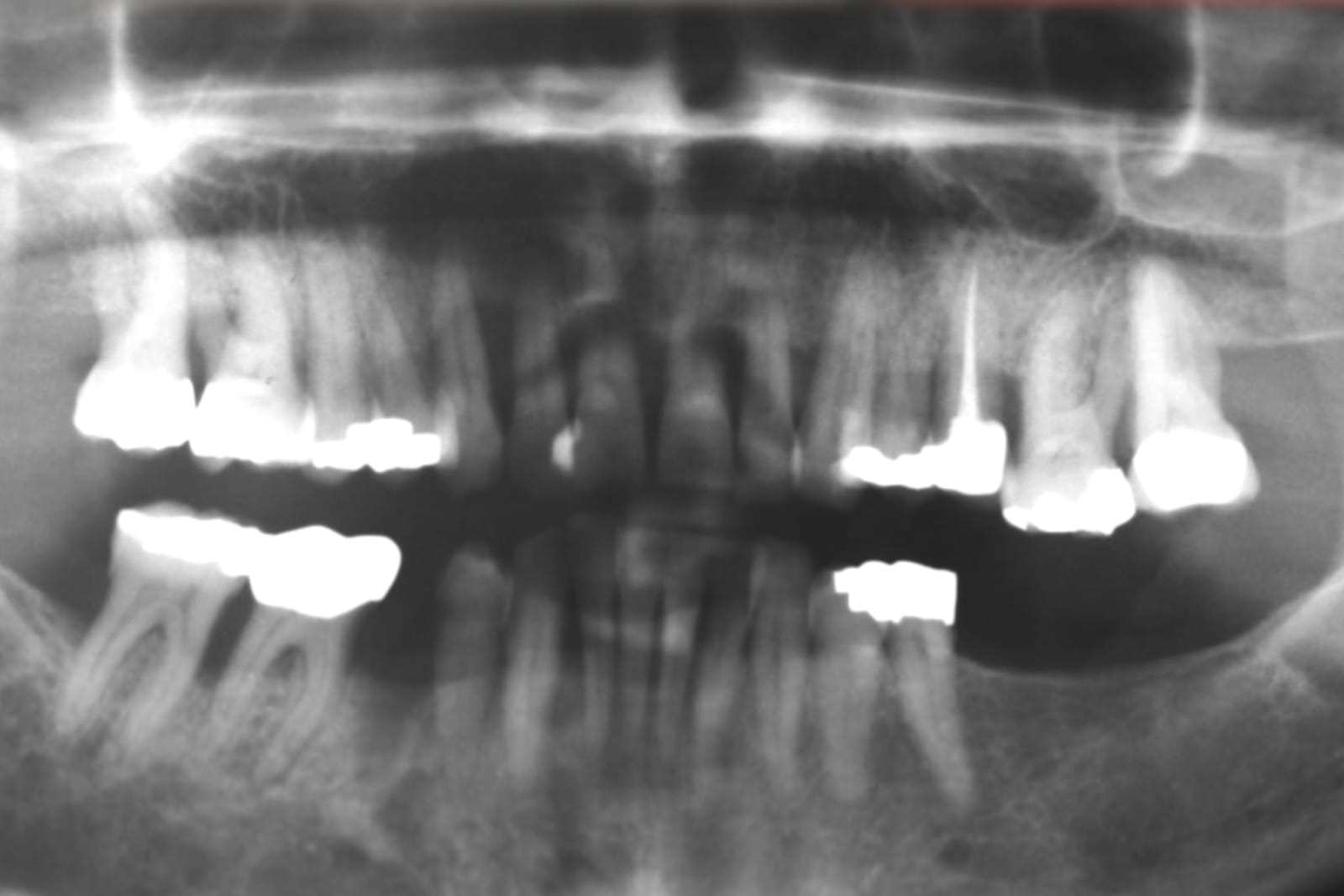
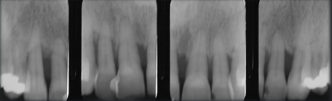
Gum disease had destroyed much of the bone around her upper teeth, especially the front ones. Removing those teeth would have let the gums and bone shrink further and left her needing an FP3 bridge with pink ceramic to replace the missing gum.
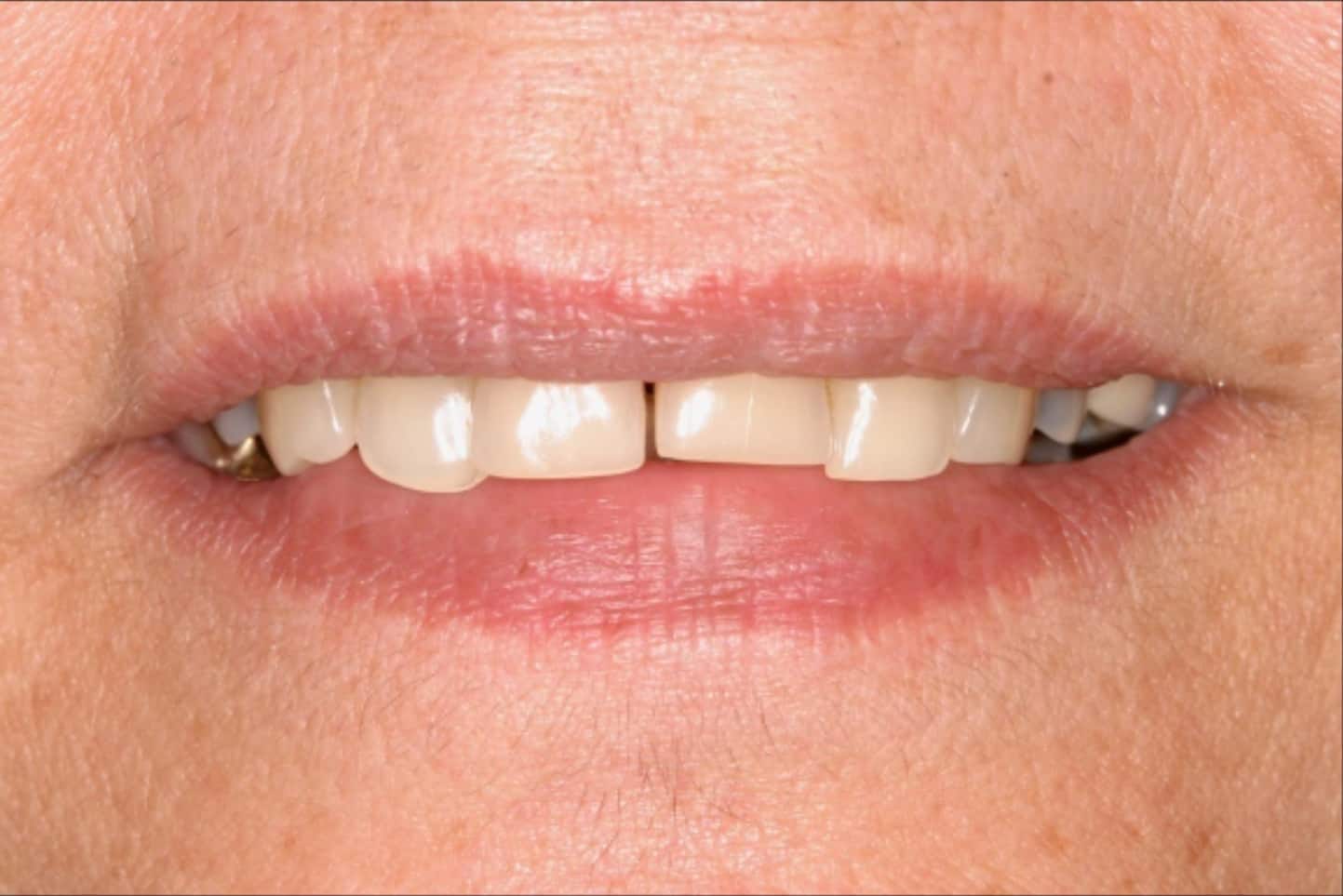
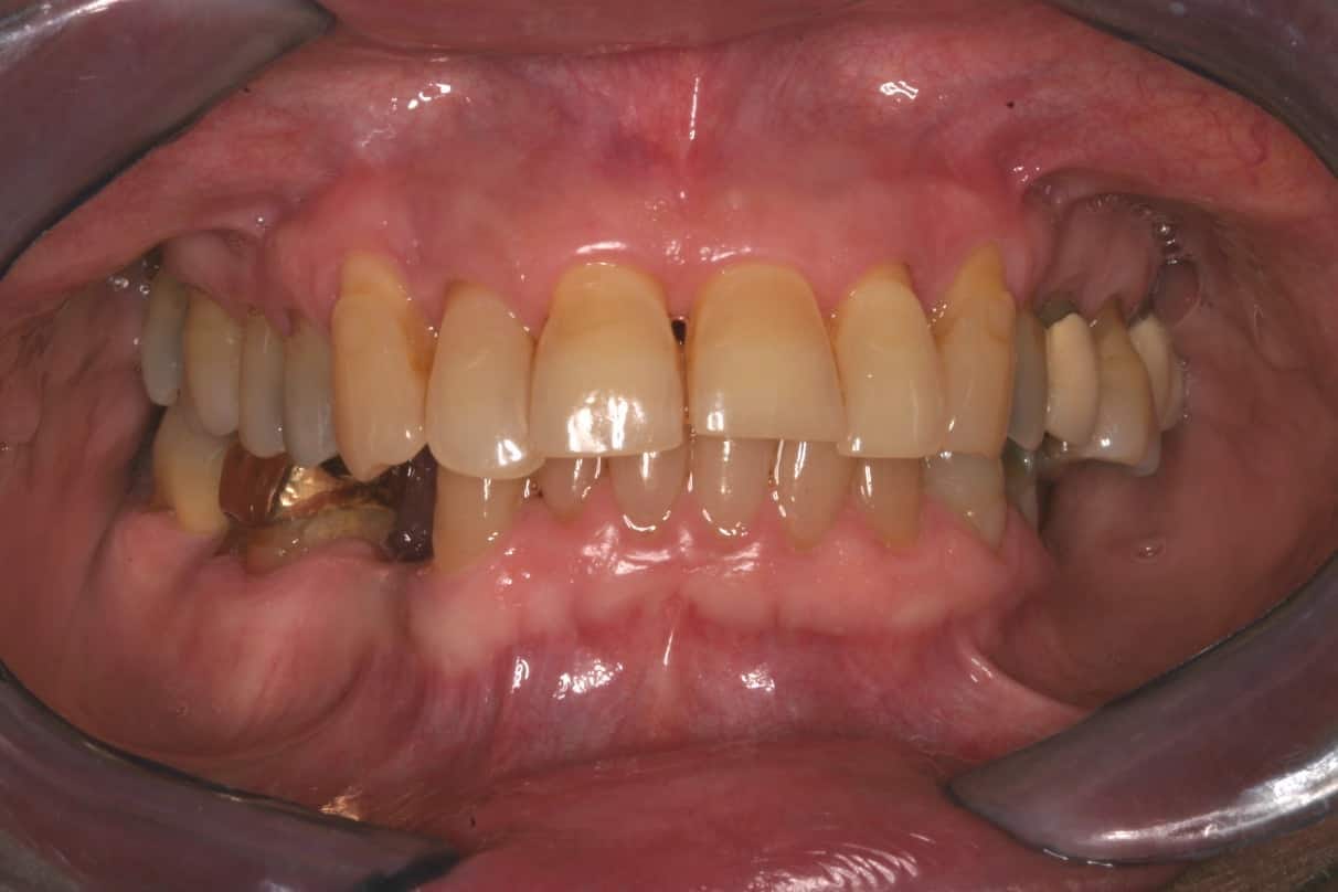
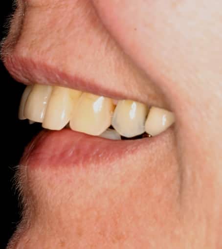
She was also missing back teeth, so her bite had collapsed. Her teeth sat at uneven, jagged levels, and with little bone to hold them, her upper front teeth had splayed forward. Her lower lip was getting caught behind them, her upper lip was pushed out, and speaking had become difficult.
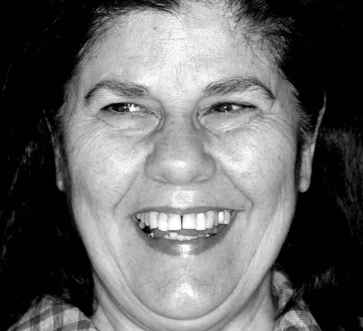
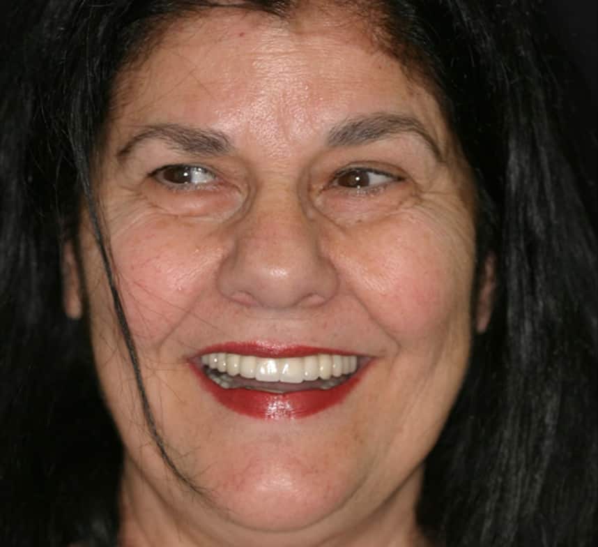
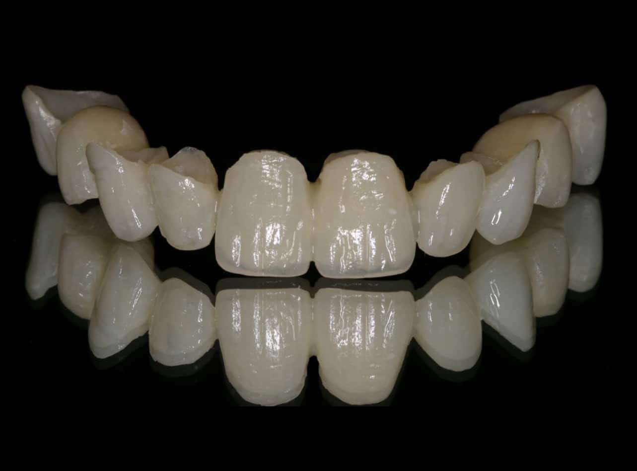
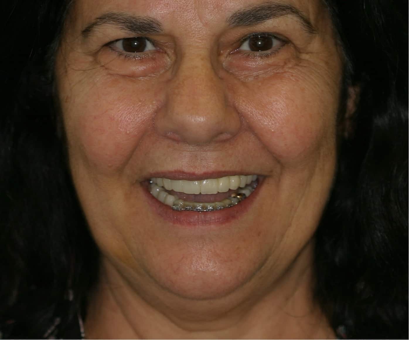
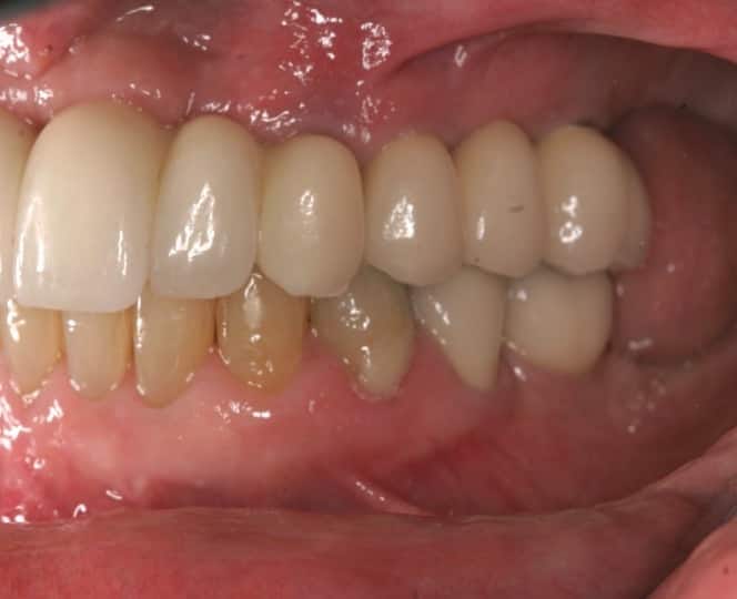
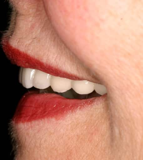
The finished ceramics
At arrival
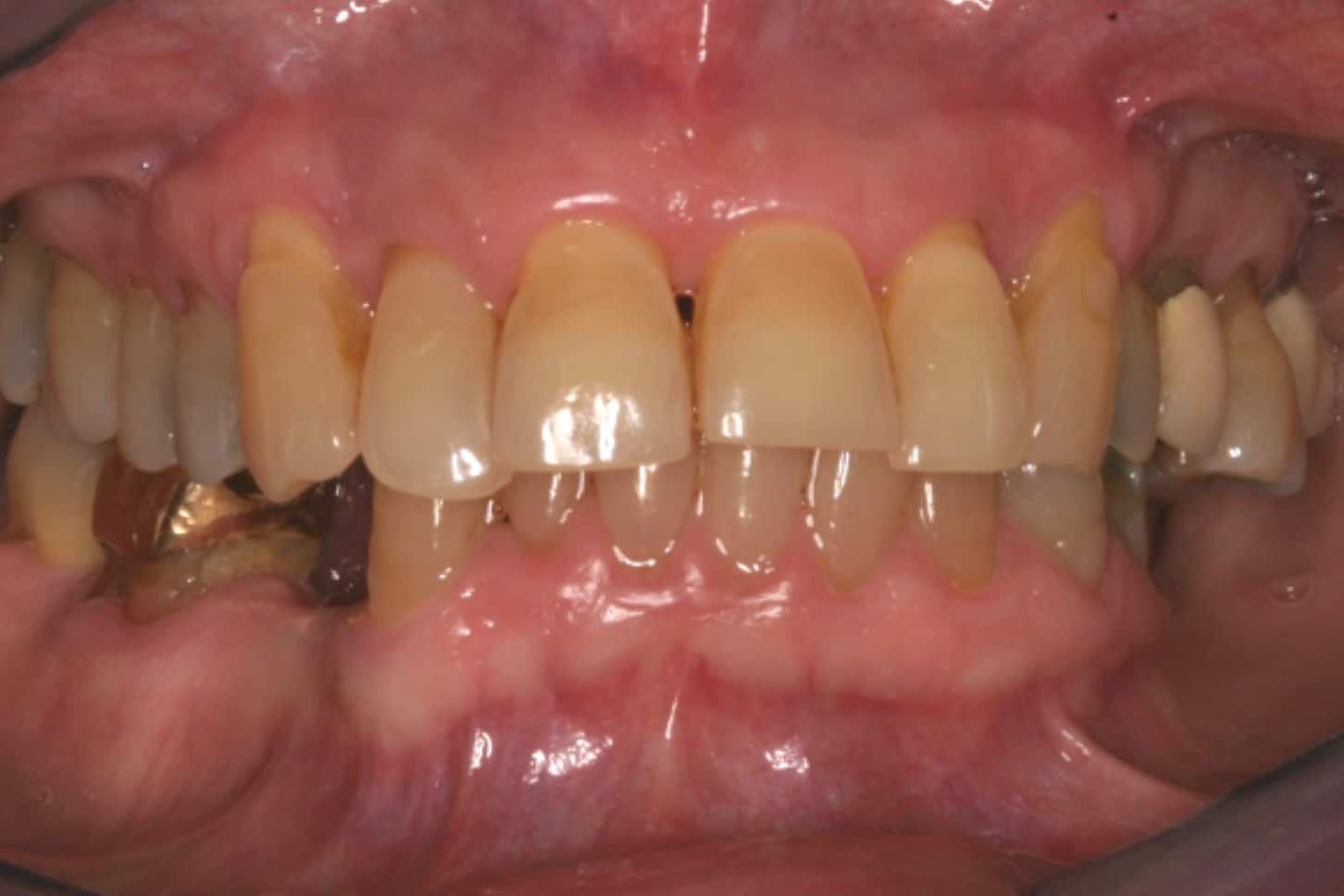
The bite and the bone
Bone around the front teeth
White ceramics only
Many clinics would remove all the teeth, cut back the gums and bone, and place an All-on-4. After talking it through with her, we chose a different goal: reverse the damage and restore her with white ceramics only.
Slow orthodontic extrusion
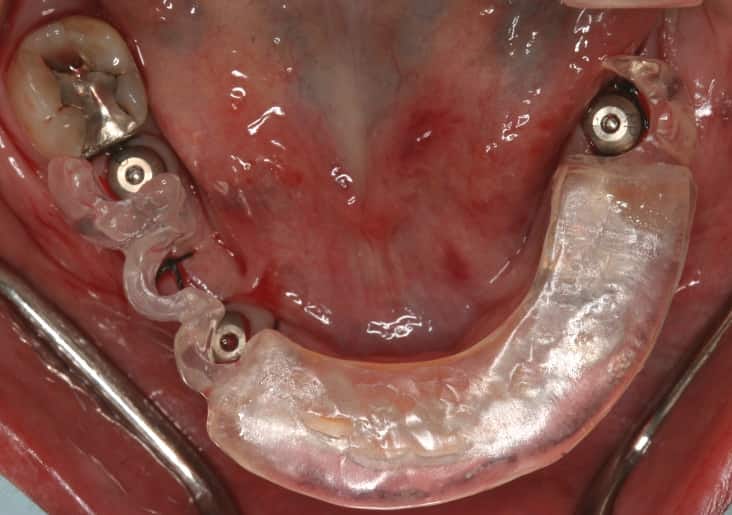
With an orthodontist, Dr. Reshad placed small anchors in the lower jaw with a wire between them. Elastics ran from that wire to the upper front teeth, so every time she opened her mouth, those teeth were drawn gently downward, bringing the gum and bone with them.
Over nine months the teeth were slowly extruded. The attachments were moved higher up the roots as the teeth came down, and the teeth were shaped as they moved. This slow orthodontic extrusion grew the bone downward, and the before-and-after X-rays show a clear gain in bone levels.
The front teeth
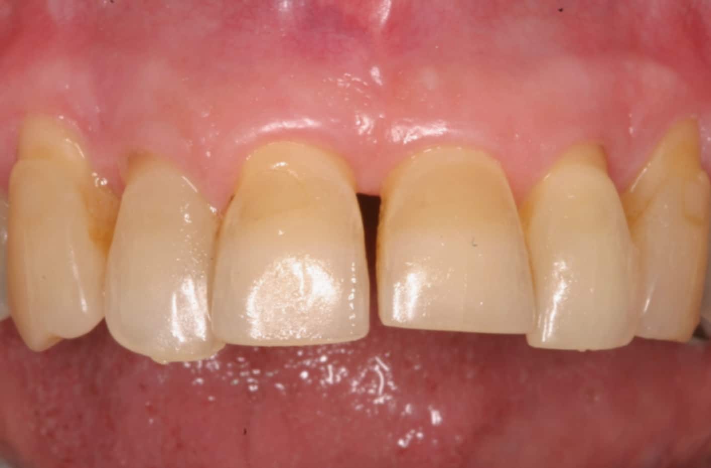
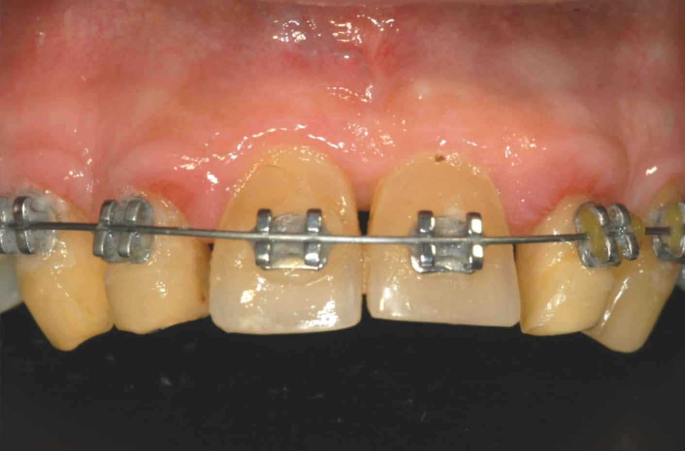
Elastics to the lower anchors
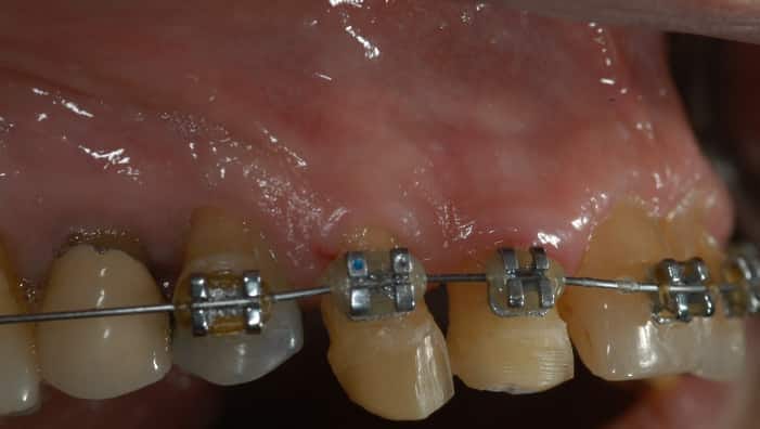
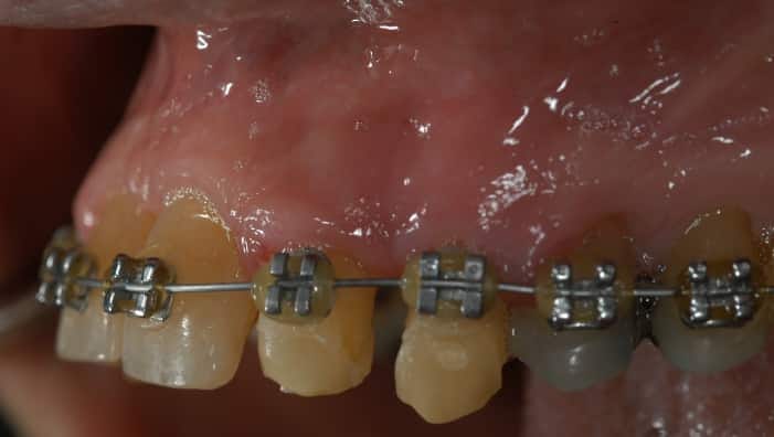
The anchor
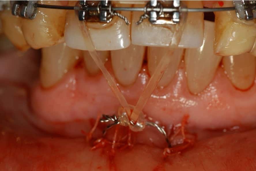
Extrusion
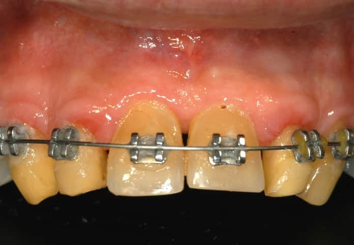
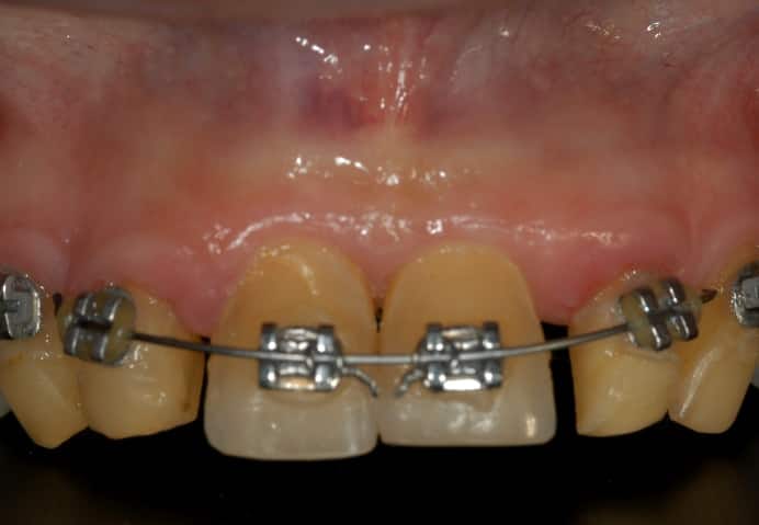
Bone levels, before and during
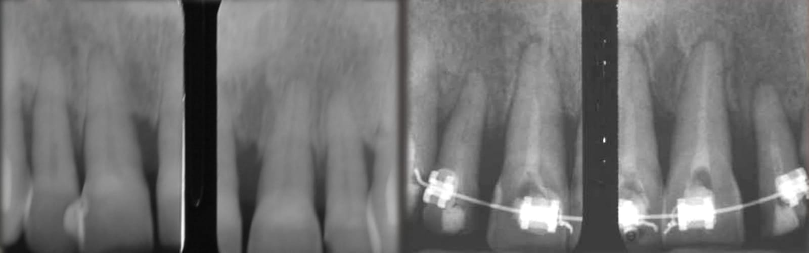
During treatment
Guided to the centre of each tooth
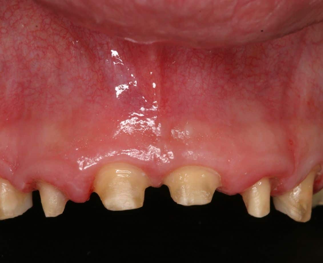
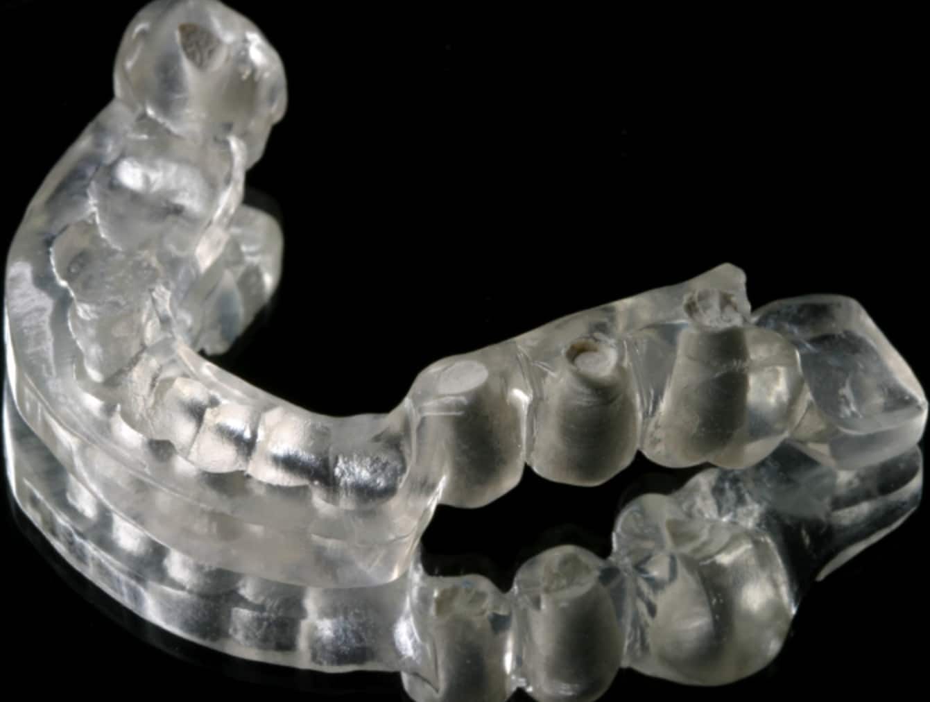
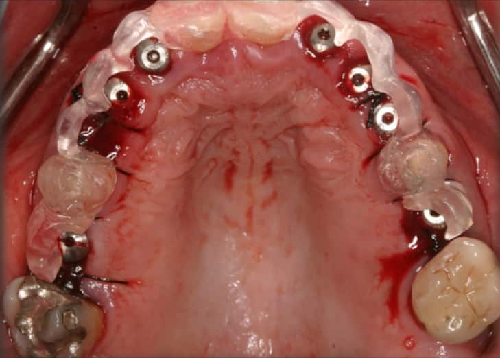
Once the bone was rebuilt, the teeth were prepared and she wore an aesthetic provisional that confirmed the gums were in the right place. Only then were implants placed, guided so each sat at the centre of its tooth, with some added grafting to reinforce the gums.
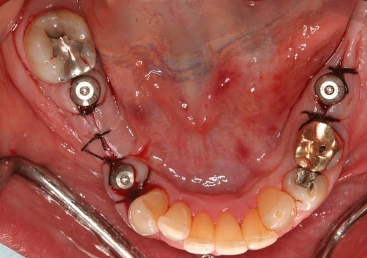
While the implants healed, the same lower anchors were used to push down her over-erupted lower front teeth, levelling the lower smile. Showing more than about 1 mm of the lower teeth is unattractive and puts the bite at risk.
Prepared, then the provisional
The surgical guide
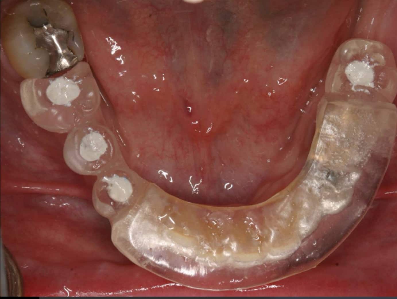
Implants placed
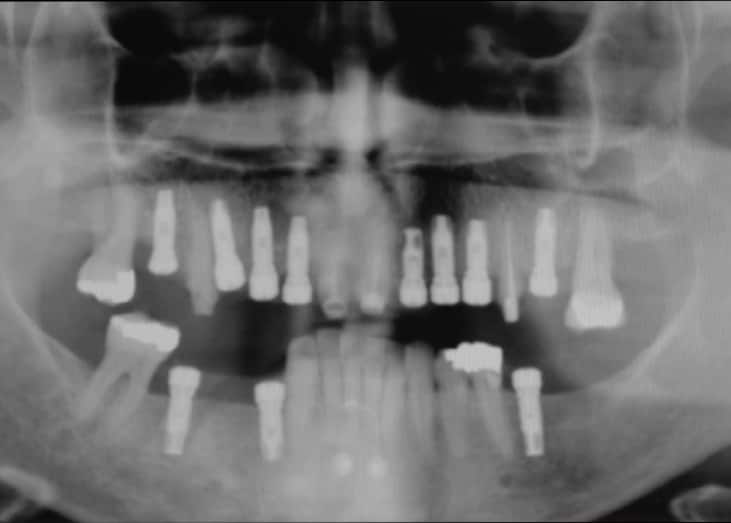
The lower anchors at work
Aesthetics, form, function and speech
With both occlusal planes corrected, a wax-up and provisionals were refined, and she received an FP1 restoration with no pink ceramic at all. That is remarkable for someone who had lost so much bone. Her side profile shows the lip support restored and the flaring gone. It is a case where aesthetics, form, function and speech all came together, led by a prosthodontist coordinating the whole team.
The upper arch, finished

Side profile
Still intact
She recently returned to Dr. Reshad's Beverly Hills office to have a single tooth treated, one he had never restored. Everything done for her about 25 years ago was still intact and looked almost exactly as it did on the day it was finished.
Every case is planned around one person's anatomy. Individual results vary, and what was right for this patient may not be right for you.